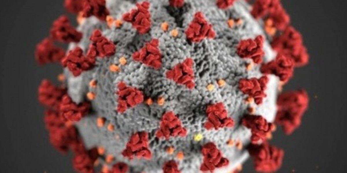
❄️? Cryo-Electron Microscopy (Cryo-EM) | Humanized Overview
Cryo-electron microscopy (Cryo-EM) is a groundbreaking imaging technology that lets scientists see biomolecules in near-atomic detail—without the need to crystallize them. Imagine being able to capture proteins, viruses, and other cellular structures in their natural, frozen state—almost like a biological snapshot in ultra-HD.
? Why It’s a Game-Changer
Cryo-EM has become a go-to tool in drug discovery, structural biology, and virology because:
It doesn’t require large crystals (unlike X-ray crystallography)
It can visualize complex and flexible proteins that are hard to study by other means
It’s especially useful for studying membrane proteins and macromolecular complexes
? How It Works (In Simple Terms)
A biomolecule sample is rapidly frozen to preserve its structure.
It’s hit with a beam of electrons inside a powerful microscope.
Thousands of 2D images are taken and digitally reconstructed into a 3D structure.
? What's Driving the Market?
? Boom in drug discovery and personalized medicine
? Rise of structural biology and genomics
? Growth of biopharma R&D
? Increasing investments from both academia and industry
⚙️ Constant improvements in AI-assisted image processing and detectors
? Challenges
? High equipment and maintenance costs
? Need for specialized data analysis tools
? Steep learning curve and lack of trained experts
? Sample prep can be tricky and time-consuming
? Applications
Cancer and neurodegenerative disease research
Infectious disease studies (e.g., visualizing SARS-CoV-2 proteins)
Vaccine and antibody development
Gene editing (e.g., CRISPR-related structures)
? Key Players
Thermo Fisher Scientific
JEOL Ltd.
Hitachi High-Tech
ZEISS
Gatan (part of AMETEK)
DENSsolutions
? Market Outlook
The Cryo-EM market is rapidly growing, with increasing adoption in both academia and pharma. It's expected to see double-digit CAGR growth, driven by its expanding use in drug discovery pipelines and structural genomics.


